Novel nanomaterial-organism hybrids with biomedical potential
Novel nanomaterial-organism hybrids with biomedical potential
Investigation on spectral and biomedical characterization of rhamnolipid from a marine associated bacterium Pseudomonas aeruginosa (DKB1)
Microbe Profile: Dictyostelium discoideum: model system for development, chemotaxis and biomedical research
The social amoeba Dictyostelium discoideum is a versatile organism that is unusual in alternating between single-celled and multi-celled forms. It possesses highly-developed systems for cell motility and chemotaxis, phagocytosis, and developmental pattern formation. As a soil amoeba growing on microorganisms, it is exposed to many potential pathogens; it thus provides fruitful ways of investigating host-pathogen interactions and is emerging as an influential model for biomedical research.
Assessment of transparency indicators across the biomedical literature: How open is open?
Recent concerns about the reproducibility of science have led to several calls for more open and transparent research practices and for the monitoring of potential improvements over time. However, with tens of thousands of new biomedical articles published per week, manually mapping and monitoring changes in transparency is unrealistic. We present an open-source, automated approach to identify 5 indicators of transparency (data sharing, code sharing, conflicts of interest disclosures, funding disclosures, and protocol registration) and apply it across the entire open access biomedical literature of 2.75 million articles on PubMed Central (PMC). Our results indicate remarkable improvements in some (e.g., conflict of interest [COI] disclosures and funding disclosures), but not other (e.g., protocol registration and code sharing) areas of transparency over time, and map transparency across fields of science, countries, journals, and publishers. This work has enabled the creation of a large, integrated, and openly available database to expedite further efforts to monitor, understand, and promote transparency and reproducibility in science.
Cryoimmunology: Opportunities and challenges in biomedical science and practice
Autologous and allogeneic cryoimmunological medicine is a new branch of biomedical science and practice that examines the features and formation of the immune response to immunogenic properties of normal and malignant biological structures altered by ultralow temperature, as well as specific changes in the structural and functional characteristics of immune cells and tissues after cryopreservation. Cryogenic protein denaturation phenomenon provides important insights into the mechanisms underlying the damage to cryogenic lesions immediately after freeze-thawing sessions in bioscience and medicine applications. The newly formed cryocoagulated protein components (cryomodified protein components) are crucial in cryoimmunology from the perspective of the formation of immunological substances at ultralow temperatures. Dendritic cells and cryocell detritus (cryocell debris) formed in living biological tissue after exposure to ultralow temperature in vivo may be an indication of one of the essential mechanisms involved in the cryoimmunological response of living structures to the impact of ultralow temperature exposure. The formation of new autologous and allogeneic cryoinduced immunogenic substances is a novel concept in biomedical science. Accordingly, this review focuses on issues concerning the peculiarities of the interaction of the immune system with a dominant malignant neoplasm tissue after exposure to subzero temperatures, considering the original cryogenic technical approaches. We present an overview of the state-of-the-art methods of cryoimmunology, and their major developments, past and present. The need for the delineation of structural and functional c


You May Also Like
Biomedical articles share annotations with their citation neighbors
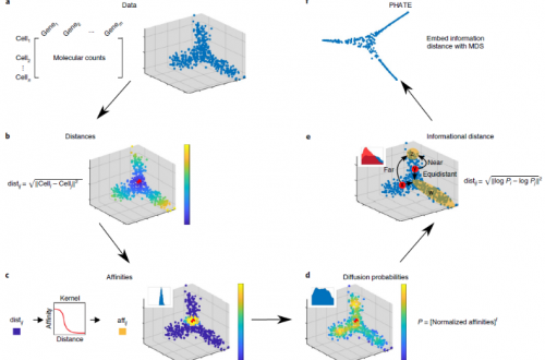
Introduction to Single Cell Sequencing Analysis (7): PHATE or Fate